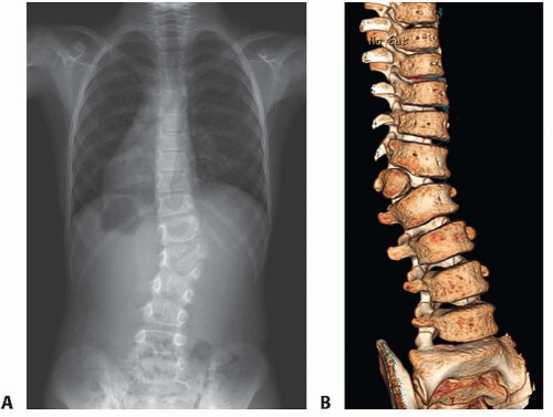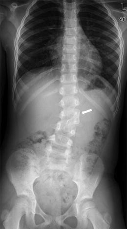


Berjano, Locarno, Switzerland (Web-publishing, Social Media) Our strategy may provide an option for similar cases with multiple consecutive cervical hemivertebrae and a large structural compensatory thoracic curve, which proved to achieve excellent correction in both the coronal and sagittal planes with acceptable neurologic risk. The neonate had multiple left lower ribs agenesis and bifid rib of right first and second ribs, dorsal left hemivertebrae in the mid and lower thoracic. The radiograph at 5-year follow-up showed that both the coronal and sagittal balance were well restored and stabilized, with the occipital tilt reduced from 12° to 0°. The cervical curve was reduced to 23° while the thoracic curve to 60° after the first-stage surgery, and the thoracic curve was further reduced to 30° after the second-stage surgery. Six months later, T6 PSO osteotomy was used to correct the structural compensatory thoracic curve. Based on our staged strategy, the three consecutive cervical hemivertebrae, as the major pathology causing the deformity, were firstly resected by the combined posterior and anterior approach. Three-dimensional computed tomography (3-D CT) scan revealed three. Three-dimensional computed tomography (3-D CT) scan revealed three semi-segmented hemivertebrae (C4, C5 and C6) on the right side. X-ray showed a right cervical curve of 60 and a left compensatory thoracic curve of 90. radiographs revealed a consistently solid arthrodesis with no evidence of curve. X-ray showed a right cervical curve of 60° and a left compensatory thoracic curve of 90°. when 2 hemivertebrae exist on opposite sides, scoliosis may be slight. The malformation developed since birth, and back pain after long-time sitting or exercise arose since 6 months before, which was unsuccessfully treated by physiotherapy. MethodsĪ 14-year-old male presented with progressive torticollis and spine deformity. In a study of more than 15,000 chest radiographs, the incidence of congenital scoliosis due to vertebral anomalies was 0.5 in 1000 livebirths (Wynne-Davies. To present our experience of staged correction with multiple cervical hemivertebra resection and thoracic pedicle subtraction osteotomy (PSO) treating a rare and complicated congenital scoliosis. When growth occurs on the affected side, it causes compression of the superior and inferior vertebral end plates, resulting in decreased growth on the concave side ( Nasca et al., 1975).Qianyu Zhuang, Jianguo Zhang, Shengru Wang, Jianwei Guo, Guixing Qiuĭecember 2015, Volume 25, Issue 1, pp 188 - 193 The abnormal vertebral body elongates the convex side of the spine. The medical significance of hemivertebrae is that they act as a wedge within the vertebral column, causing a curvature away from the side of the defect ( Zelop et al., 1993). Clinically this results in a kyphosis rather than a scoliosis. This latter defect occurs when the anterior part of the vertebral body fails to develop. Secondary degenerative disc disease can lead to increase kyphosis. (1975) classified hemivertebrae by their morphologic appearance and described six different types: single supernumerary hemivertebrae, single wedge-shaped hemivertebrae, multiple hemivertebrae, multiple hemivertebrae with a unilateral bar defect on the contralateral side, balanced hemivertebrae, and posterior hemivertebrae. Abnormalities in vertebral segmentation result in bar or block vertebrae, whereas abnormalities in formation result in hemivertebrae ( McMaster and Ohtsuka, 1982). Abnormalities of the vertebral bodies result from either failure of formation or failure of segmentation ( Abrams and Filly, 1985). These centers fuse, resulting in three primary centers of ossification, which can be visualized sonographically as early as 12 weeks of gestation, but histologically as early as 8 weeks of gestation ( Zelop et al., 1993). Each vertebral body has a dorsal and ventral primary ossification center. Bidimensional and 3-4D ultrasound imaging as well as radiograph of the fetus and. At approximately 6 weeks of gestation, a chondrification center appears for each mesenchymal vertebra. Ultrasound showed spine deformation, hemivertebrae and crab-like ribs. Once the mesenchymal pattern is established in the embryo, subsequent cartilaginous and osseous stages follow ( McMaster and Ohtsuka, 1982). Hemivertebrae and fusions in the lower thoracic and lumbar areas may present as an incidental finding on radiography, single curve scoliosis, or as pain. Plain radiograph / CT Usually directly outlines the bony anomaly and is often seen as a wedge-shaped vertebral body. These anomalies develop during the first 6 weeks of gestation, when the future anatomical pattern of the spine is formed in mesenchyme. Radiographic features Antenatal ultrasound A hemivertebra may be seen as an asymmetrical vertebral body on sagittal or coronal scanning, while on axial scanning, a focal defect may be seen on either side of the vertebral column 5. Hemivertebrae are vertebral anomalies that can be detected sonographically by the second trimester of pregnancy.


 0 kommentar(er)
